Para-Buster
Showing posts with label Alzheimer's. Show all posts

Saturday, March 29, 2008

With Alzheimer's, It Takes a Family

(HealthDay News) -- Alzheimer's care in the United States is a family affair, a new survey suggests.

Three in five so-called "sandwich" caregivers who look after a relative or friend with Alzheimer's disease said their children help with care duties, ranging from attending doctors' appointments to feeding and dressing their loved ones, according to the third annual Alzheimer's Foundation of America Investigating Caregivers' Attitudes and Needs Survey.

Sandwich caregivers are parents or guardians of children under age 21 who also care for an aging parent, relative or friend with Alzheimer's.

The survey of 559 sandwich caregivers found that among those who feel they do a good job balancing their care of a loved one with Alzheimer's and looking after their children, 36 percent said support from their children is a factor in their success.

Among children aged 8 to 21 who are involved in caregiving:

  • about one-third of young adults (aged 18-21) help with doctors' appointments;

  • 42 percent of young adults assist with transporting loved ones with Alzheimer's disease;

  • about one-quarter of young adults and teens (aged 13-17) help with the activities of daily living, such as feeding and dressing;

  • nearly 90 percent of pre-teens (aged 8 to 12) visit and entertain a loved one with Alzheimer's disease;

  • about 85 percent of teens visit loved ones with the disease.


"Taking care of someone with Alzheimer's disease can be an enormous drain on the caregiver and on family resources. For sandwich caregivers, the problem is even more acute. It is clear that caregiving is a multigenerational concern. Young adults, and even teens and pre-teens, are being impacted in life-changing ways by their caregiving responsibilities," Eric J. Hall, president and chief executive officer of Alzheimer's Foundation of America, said in a prepared statement.

The survey also found that 70 percent of sandwich caregivers said they need more help caring for a loved one with Alzheimer's, while 33 percent said they need more help with their children. In addition, 63 percent said they'd like more information about how to help their children cope when a loved one is diagnosed with Alzheimer's disease.

"A segment of young adults and teens assist with managing the daily needs of individuals with Alzheimer's disease, and a small percent are even called upon to make informed decisions about treatment. It's crucial that they have access to good information sources," Dr. Lesley Blake, a clinical associate professor of psychiatry at the University of Washington School of Medicine in Seattle, said in a prepared statement.

"As Alzheimer's disease progresses, declines in cognition, function and behavior worsen. Both adult and non-adult caregivers need to be educated about what to expect and, more importantly, what to do in these cases," Blake said. "Proper diagnosis and treatment are crucial. Symptoms -- loss of function, decline in cognitive ability and difficult behavior -- can be delayed and caregiver burden reduced through medication therapy, which may include combining medications from two FDA-approved Alzheimer's medication classes."

The survey, funded by Forest Pharmaceuticals Inc., found that 77 percent of sandwich caregivers weren't aware that combination drug therapy can be used to treat Alzheimer's disease. In many cases, respondents said there was a delay -- typically about two years -- in their loved ones' diagnosis of Alzheimer's. In cases where diagnosis was delayed for a year or more, the most common cause was a lack of caregiver awareness about Alzheimer's disease. About half of those caregivers said they believed the disease was a normal part of aging.

It's estimated that more than 5 million Americans have Alzheimer's disease -- about one in 10 of those aged 65 and older and nearly half those 85 or older -- and that number could more than triple, to 16 million, by 2050, according to background information in a news release about the survey.

More information
The U.S. National Institute on Aging has more about caring for someone with Alzheimer's.

Monday, January 28, 2008

Alzheimers Research Target May Be a Dead End

(HealthDay News) -- A once-promising pathway for research into preventing and treating Alzheimer's disease may have been derailed by a surprise chemical finding, researchers report.

Scientists in laboratories around the world have been investigating drug candidates called amyloid inhibitors, which many experts believed could keep proteins such as amyloid-beta from sticking together in brain tissue.

This type of "sticky" protein plaque build-up is a hallmark of Alzheimer's disease. It also characterizes brain illnesses such as Huntington's disease and "mad cow" disease.

But the new study, published Jan. 27 in the journal Nature Chemical Biology, may sound an unexpected death knell for amyloid inhibitor research.

In the study, a team of chemists at the University of California, San Francisco, found that these candidate drugs form large, unwieldy clumps themselves, rendering them useless as targeted therapy against amyloid in the brain.

High-tech research in the lab is revealing that typical amyloid inhibitors "seem to act not in the way people expect them to and want them to," explained study senior author Brian Shoichet, professor of pharmaceutical chemistry at UCSF.

Once these drugs aggregate into clumps, "they no longer have the right pharmacology, they won't cross the [brain's] membrane barriers, and they inhibit everything -- any protein will bind with them," he said.

In other words, the drugs lose their ability to migrate to the brain to fight amyloid plaque. They also give up their targeted specificity against amyloid, Shoichet said. "They end up inhibiting everything -- any protein that sees them will be sequestered by them," he said. This molecular clumping process is largely inevitable, Shoichet added.

His advice to neuroscientists investigating these agents as potential Alzheimer's therapies: "They should stop."

Another expert agreed.

David Lynn is a Howard Hughes Institute investigator and professor of biological chemistry at Emory University in Atlanta. "I think that Brian's paper argues that [scientists] have been missing the boat here," he said. "It's not clear that you are ever going to get the concentrations that you need of these agents at the right site to be able to have any therapeutic intervention."
On the level of basic chemistry, attacking Alzheimer's and other protein-clumping diseases by preventing amyloid from concentrating has "always been a long shot," Lynn said. That's because amyloid proteins are incredibly "sticky," chemically speaking.

"To find things that will competitively stick and stop them from assembling is theoretically hard to imagine," Lynn said. It was thought that individual molecules of amyloid inhibitors might do so, but the new finding -- that the molecules inevitably bind together in a more impractical mass -- renders them therapeutically useless.

But other avenues of Alzheimer's research remain promising, Lynn said.

"There are certainly other strategies that have potential," he noted, including antibody-focused strategies aimed at eliminating plaques, or treatments focused on easing the downstream effects of amyloid buildup.

Both scientists stressed that it's still not certain whether protein plaques even cause Alzheimer's and other brain diseases, or whether they are merely byproducts of the disease process. "That's really another open area of research," Shoichet said.

"The problem with these diseases is that it is such a moving target," Lynn said. "And so, different people are looking at different things."

More information
There's much more on Alzheimer's disease at the Alzheimer's Association.

Friday, October 19, 2007

Tooth Troubles Could Raise Dementia Risk

(HealthDay News) -- Tooth loss and mouth illnesses may boost the risk of dementia later in life, U.S. research shows.

A team at the University of Kentucky analyzed the dental records and annual cognitive test results of 144 participants, ages 75 to 98, in the Nun Study, an examination of aging and Alzheimer's disease among sisters of the School Sisters of Notre Dame.

Of participants "who did not have dementia at the first examination (of annual exams over a 12-year period), those with few teeth -- zero to nine -- had an increased risk of developing dementia during the study, compared with those who had 10 or more teeth," the study authors wrote in the October issue of the Journal of the American Dental Association.

The team offered several possible reasons for this association, including periodontal disease, early-life nutritional deficiencies, and infections or chronic diseases that may result simultaneously in tooth loss and brain damage.

Further research is needed to confirm whether there is a direct link between tooth loss and increased risk of dementia, the researchers said.

"It is not clear from our findings whether the association is causal or casual," they wrote.

More information
The U.S. National Institute of Neurological Disorders and Stroke has more about dementia.

Tuesday, July 10, 2007

Dementia Gene Mutation Identified

(HealthDay News) -- Researchers have discovered a new gene mutation linked to frontotemporal dementia, a disease that affects language abilities and socially appropriate behavior in the people who have it.

Frontotemporal dementia is the second most common form of dementia after Alzheimer's disease.

After a decade of work on the question, researchers at the Regional Neurogenetic Centre in Lamezia Terme, Italy, and the Centre for Research in Neurodegenerative Diseases at the University of Toronto identified the mutation in a gene named progranulin by studying the genealogy of 15 generations of an extended Italian family. Thirty-six members of the family have had frontotemporal dementia. Researchers conducted DNA tests on 70 family members, including 13 with the disease.

The mutation is in a gene on chromosome 17, which leads to a loss of progranulin, a protein growth factor that helps brain cells survive. The mutation limits production of the protein to half the normal amount, because only one copy of the gene is active. While reduced production of progranulin is related to dementia, an excess has been tied to cancer.

The researchers reported finding the mutation in nine of the family members with the disease and in 10 people who were too young to have the disease. They noted that four people who have the disease did not have the mutation, but were descendants of a line of the family in which three generations had frontotemporal dementia, indicating a second possible genetic link.

They also noted that the age at which people with the mutation began to see symptoms of frontotemporal dementia varied between 35 and 78 years old.

The findings are published in the July 10 issue of Neurology.

More information
To learn about frontotemporal dementia, visit the National Institute of Neurological Disorders and Stroke.

Tuesday, June 12, 2007

New Techniques Could Spot Alzheimer's Early

(HealthDay News) -- Three groups of researchers are reporting progress on the early detection of Alzheimer's disease -- advances that, if validated, could aid patients and drug developers alike, experts said.

In one study, a Norwegian team identified a gene expression "signature" that distinguishes people with Alzheimer's from healthy individuals with an up-to-85 percent accuracy.
In other research, U.S. scientists reported that a combination of brain imaging techniques with sophisticated "pattern recognition software" could differentiate those with mild cognitive impairment (an early form of Alzheimer's) from those without the condition with 100 percent accuracy.

Finally, another U.S. study reveals that key diagnostic algorithms can predict an individual's six-year risk of developing dementia with at least 87 percent accuracy.

The results were announced Sunday at the Alzheimer's Association's International Conference on Prevention of Dementia in Washington, D.C.

There currently is no cure for Alzheimer's disease, which the Alzheimer's Association now estimates affects nearly five million Americans. Existing treatments can slow progression of the brain-wasting disease, but by the time an individual has been diagnosed the disease has typically been at work for years and extensive, irreparable tissue damage has already occurred.

Diagnosis is currently done simply by observing patients and is only about 80 percent accurate.
According to Greg Cole, associate director of research for the Geriatric Research Education and Clinical Center at the Greater Los Angeles Veterans Administration Medical Center, early detection of Alzheimer's disease can aid both patients and those scientists searching for a cure.

Patients benefit, he says, because earlier treatment can minimize tissue loss and thus improve quality of life.

"It's like termite damage," he said. "You want to repair the damage before they destroy your house, you will be so much better off."

Pharmaceutical companies, on the other hand, can more confidently identify affected populations, which can reduce the size and cost of clinical trials, and thus enable them to perform more of them.

The Norwegian study, led by Anders Lonneborg, research director at DiaGenic ASA in Oslo, focused on gene expression profiling of an individual's blood.

Using a 1,200-gene DNA microarray, his team could distinguish those with Alzheimer's disease from those who were healthy with 85 percent accuracy. Using a smaller set of genes (fewer than 96) and a technique called RT-PCR, the team achieved an accuracy of 79.5 percent.

But that was using samples from individuals whose disease was so far advanced that they could be diagnosed clinically, anyway. It remains to be seen, Cole said, if the test can pick out individuals much earlier in the disease process, when treatment might be more effective.

That, Lonneborg added, is the next step. "Now we will test [the diagnostic] on risk groups who have minor memory problems and see if we can predict who will develop Alzheimer's disease in a few years. This will be very interesting to see how it works," he said.

A second study tackled early diagnosis using sophisticated brain imaging. The technique was developed by a team led by Christos Davatzikos, of the department of radiology at the University of Pennsylvania in Philadelphia.

Combining MRIs of brain structure with positron emission tomography (PET) measurements of cerebral blood flow, the new test uses pattern recognition software to identify spatial patterns that pinpoint with 100 percent accuracy whether an individual does or does not have mild cognitive impairment.

Using data from the Baltimore Longitudinal Study of Aging, Davatzikos' team gained access to individual patients' brain images and blood work from over a period of years.

"We found that even before the most subtle clinical symptoms, when they were healthy, we were able to see abnormality scores that were indicative of mild cognitive impairment in at least half of them," said Davatzikos. "So there were early signs."

Cole applauded this test's accuracy, but noted that it is unlikely to have wide application because of its cost -- several thousand dollars. Davatzikos said his team is now working to balance the test's accuracy and cost, for instance, by optimizing the accuracy of MRI-based tests alone.
Finally, a team led by Deborah Barnes, assistant professor of psychiatry at the University of California, San Francisco, is using a special "bedside" algorithm, or formula, to try and predict an individual's dementia risk.

This tool -- akin to similar methods for calculating the risk of cardiovascular disease or diabetes -- predicts with up to 88 percent accuracy whether an individual will develop dementia over the next six years.

Based on such variables as age, cognitive function and physical performance, the index is an inexpensive, fast and practical algorithm that groups individuals into low-, medium-, and high-risk categories. Those in the low-risk group have a 6 percent risk of developing dementia, compared to 54 percent for those in the high-risk category.

According to Barnes, such an index can provide comfort to those least likely to develop disease while motivating those at higher risk to adopt a healthier lifestyle.

At the same time, it provides information so their families may begin planning for whatever the future may bring, she added.

More information
For more information on Alzheimer's Disease, visit the Alzheimer's Association.

Saturday, January 13, 2007

A Comprehensive Review of Heavy Metal Detoxification and Clinical Pearls from 30 years of Medical Practice by Dietrich Klinghardt, MD.,Ph.D

A. Introduction:
Heavy Metals appear in the mammalian system because they have become part of our environment.

We are in a constant exchange with our environment which is goverened by the laws of osmosis.

If mercury is in the fish we eat, over time we have mercury in our system. We cannot keep our system pristine and clean, because we are seperated from our toxic environment only by semi-permeable membranes: skin and mucosal surfaces.

Maintaining relative cleanliness requires a number of inherentdetox systems to work overtime against the osmotic pressure of the incoming toxins. As the toxixity of our environment increases so does the osmotic pressure, pushing the often man- made poisons into our body.

Toxins almost never come alone. They come in synergistically acting package-deals. Mercury alone is toxic. Together with zinc it is many times more toxic, add in a little copper and silver, as in dental amalgam fillings and the detrimental effect to the body increases manyfold.

Together with mercaptan and thioether (dental toxins) the toxic amalgam effects grow exponentially.

Add in a little PCB and dioxin, as in fish, and the illness causing effect of the methyl mercury in fish increases manyfold. Toxicology is to a large degree the study of synergistic effects.

In synergy 1 plus 1 = 100. Heavy metals are primarily neurotoxins. There is a synergistic effect between all neurotoxins which is responsible for the illness producing effect.

Making the neurotoxin elimination a major part of my practice has been an amazing experience.

Many illnesses considered intractable respond when the related issues are successfully resolved.

What are Neurotoxins?

Neurotoxins are substances attracted to the mammalian nervous system. They are absorbed by nerve endings and travel inside the neuron to the cell body. On their way they distrupt vital functions of the nerve cell, such as axonal transport of nutrients, mitochondrial respiration and proper DNA transcription. The body is constantly trying to eliminate neurotoxins via the available exit routes: the liver, kidney, skin and exhaled air.

Detox mechanisms include acetylation, sulfation, glucuronidation, oxidation and others. The liver is most important in these processes. Here most elimination products are expelled with the bile into the small intestine and should leave the body via the digestive tract. However, because of the lipophilic/neurotropic nature of the neurotoxins, most are reabsorbed by the abundant nerve endings of the enteric nervous system (ENS) in the intestinal wall. The ENS has more neurons than the spinal chord. From the moment of mucosal uptake the toxins can potentially take 4 different paths:

1. Neuronal uptake and via axonal transport to the spinal chord (sympathetic neurons) or brainstem (parasympathetics) – from here back to the brain.
2. Venous uptake and via the portal vein back to the liver
3. Lymphatic uptake and via the thoracic duct to the subclavian vein
4. Uptake by bowel bacteria and tissues of the intestinal tract

i) Heavy metals:
mercury, lead, cadmium, iron, manganese and aluminum (are the most common).Common Sources: metallic mercury vapor escapes from dental amalgam fillings (they contain about 50% mercury, the rest is zinc, silver copper, tin and trace metals). Cadmium: car fumes, cigarette smoke , pigment in oil paint Lead: outasing from-paint, residues in earth and food chain from time when lead was used in gasoline, contaminated drinking water Aluminum: cookware, drinking water

ii) Biotoxins:
such as tetanus toxin, botulinum toxin (botox), ascaridin (from intestinal parasites), unspecified toxins from streptococci, staphylococci, lyme disease, clamydia, tuberculosis, fungal toxins and toxins produced by viruses. Biotoxins are minute molecules (200-1000 kilodaltons) containing nitrogen and sulfur. They belong to a group of chemical messengers which microorganisms use to control the host´s immune system, host behaviour and the host´s eating habits.

iii) Xenobiotics (man-made environmental toxins):
such as dioxin, formaldehyde, insecticides, wood preservatives, PCBs etc.
iv) Food Preservatives, excitotoxins and cosmetics:
aspartame (diet sweeteners), MSG, many spices, food colourings, fluoride, methyl-andpropyl -paraben, etc.

Heavy Metal Toxicity
Metals can exist in the body with different kinds of chemical bonds and as different molecules. Mercury appears to be the king-pin in the cascade of events in which metals become pathogenic. Mercury can be present as metallic mercury (HgO), as mercury salt (e.g. mercury chloride – HG+), or as methyl mercury (HG++). Methyl mercury is 50 times more toxic than metallic mercury. Methyl-Hg is so firmly bound to the body that it has to be first reduced to HG+ before it can be removed from the cell. This is achieved with reducing agents (“antioxidants”) e.g. intravenous vitamin C and reduced glutathione.

To remove Hg-Salts or metallic Hg from the outside of the cell, other agents are useful Mercury belongs to a group of metals that oxidize in the presence of sulfur and form compounds with sulfur (sulfhydryl affinitive metals). Methyl mercury is already oxidized to its maximum and bound firmly to sulfur in the different proteins of the body. The following metals belong to the sulfhydryl affinitive group and respond to similar detoxification methods: Copper, arsenic, cadmium, lead, mercury. Aluminum and iron for example would not respond a sulfur compound. Some detox agents have multiple mechanisms by which they bind to metals. The algal organism chlorella has over 20 known such mechanisms.

Other metals oxidize with oxygen. Iron turns to rust when oxidized. Rust is nontoxic to the body, whereas iron is. Iron overdose responds to a chemical called desferoximin (desferal). Aluminum responds to the same detoxification agent. A recent Japanese study showed that Chinese parsley, cilantro, is a powerful elimination agent for aluminum stored in bone and the brain.

Other facts:
· Some metals are extremely toxic, even in the most minute dose, whereas others have very low toxicity, even in high doses. However, dependent on the dose, all metals can become toxic to the body. Iron can cause severe oxidative damage, copper may compromise liver function and visual acuity, selenium and arsenic have been known to be used to murder people and so on.
· Most metals serve a functional role in the body. For example, selenium is needed in the enzyme that restores oxidized glutathione back to its functional form as reduced glutathione. Another important function of selenium is its role as a powerful antioxidant in preventing cancer.
· Some metals have a narrow physiological range. That means the difference between a therapeutic dose and toxic overdose is very small. Selenium is an example of this. Magnesium on the other hand has a wide physiological range and thus is more difficult to overdose.
· Some metals have no physiological function. Mercury, lead, aluminum are in this group. Even the smallest amounts have negative physiological-effects.
· biochemical individuality: some people may react more or less than others to the presence of heavy metals in the tissues. Some people may develop a severe chronic illness after exposure of a few molecules of mercury, whereas others may be more resistant to it. Genetic deficiencies in the enzymes responsible for the formation of the metallothioneins and glutathione production and reduction are examples.

Possible side-effects during heavy metal detox:
Every patient can be affected by metals in two ways:
1. Through their non-specific toxic effects
2. Through the system´s allergic reactions to the neurotoxins
Often these two distinctive types of symptoms cannot be easily distinguished. During a detox program, the patient may also temporarily become allergic to the various substances that help to carry out the toxins. This is based on a physiological mechanism called ‘operant conditioning’.

Every time the detoxifying substance is given, mercury emerges from its hiding places into the more superficial tissues of the body, where mercury can now be detected by the immune system. The immune system however is fooled into thinking that the detoxifying substance itself is the enemy. The immune system now starts to react to the detoxifying substance as if it was the mercury itself. This reaction typically resolves spontaneously after six weeks of not using the detox agent in question. This type of conditioned reflex can also be easily treated with simple techniques e.g. NAET, PK (APN), or by giving the detox substance in a homeopathic dilution for a few days. Often the basal membranes in the kidney will swell as a sign of the allergic reaction, causing low back pain, anuria or inability to concentrate urine.

Neuraltherapy or microcurrent stimulation of the kidneys quickly resolves the issue. Muscle aches indicate the redistribution of toxins into the connective tissue and an insufficient program. Depression, headaches, trigeminal neuralgia, seizures, increased pain levels indicate redistribution of metals into the CNS and an inappropriate detox program. Eye problems and tinnitus that occurs during detox indicates redistribution of metals into these organs and requires selective mobilization from these locations before the program is continued. I use a specific type of microcurrent for this purpose

Some recently published findings related to the metal issue:
Iron/mangnese: A recent paper on Parkinsons disease (Neurology June 10, 2003;60:1761-1766)revealedthat just by eating iron and manganese containing foods such as spinach or taking supplements containing Mn or Fe - the risk of developing PD increased almost 2 fold. This demonstrates that even dietary supplements or organically grown foods are amongst the possilbe culprits in metal toxixity.

Methylmercury:
There are two major sources:
1. mercury escaped from dental amalgam fillings is converted by oral and intestinal bacteria to methylmercury, which then is bound firmly to proteins and other molecules. Methyl mercury crosses the blodd brain barrier and the placental barrier leading to massive prenatal exposure. Earlier studies determined that over 90% of the common body burdon of Hg is from dental fillings. Recent studies show that eating fish is starting to compete with amalgam fillings for the leading position as a risk factor.
2.Seafood
A recent study (JAMA, April 2, 2003;289(13):1667-1674) revealed the following It is estimated that nearly 60,000 children each year are born at risk for neurological problems due to methylmercury exposure in the womb. One in 12 U.S. women of childbearing age have potentially hazardous levels of mercury in their blood as a result of consuming fish, according to government scientists. The U.S.FDA recommends that pregnant women and those who may become pregnant avoid eating shark, swordfish, king mackerel, and tile fish known to contain elevated levels of methylmercury, an organic form of mercury. Nearly all fish contain some amount of methylmercury. Mercury accumulates in the system, so larger, longer-lived fish like shark or swordfish contain the highest amounts of mercury and pose the largest threat if eaten regularly.
The National Center for Policy Research for Women & Families published in May 2003, that the following fish are lowest in methyl mercury:
· Catfish (farmed)
· Blue Crab (mid-Atlantic)
· Croaker
· Fish Sticks
· Flounder (summer)
· Haddock
· Trout (farmed)
· Shrimp
The FDA also recommends these fish as safe to eat:
haddock, tilapia, wild alaskan salmon,and sole

Ethylmercury:
A recent quote from Boyd Haley, PhD: “our latest research clearly points to the ethylmercury exposureas being causal in autism. The tremendous enhancement of thimerosal toxicity by testosterone and the reduction of toxicity by estrogen explains the fact that 4 boys to 1 girl getting the disease and the fact thatthe bulk of severe autistics are boys. Most importantly, this autistic situation clearly shows that exposureto levels of mercury that many "experts" considered safe was capable of causing an epidemic of a neurological disease”.

B. Symptoms
Other authors have tried to specify typical symptoms for each metal. Because of the synergistic effects and simultaneous occurence of several toxins at the same time. The best source of literature on the effects of specific metals on the system are the old homeopathic textbooks ‘materia medica’ (Kent, Boericke).

I prefer to look at a client in a systemic way, not focussing on single issues . A manganese typical symptom (ie violent behaviour) may be a lot more worrysome in a given patient then their particular mercury related symptom (ie insomnia). However, the practical focus of detox should be almost always on the mercury first. If mercury is adressed appropriately, the manganese often leaves the body as a side effect of mercury detox. The opposite is not true.
Any illness can be caused by, or contributed to, or exagerated by neurotoxins. Here is a short list:
· Neurological problems: Fatigue, depression, insomnia, memory loss, blunting of the senses, chronic intractable pain (migraine, sciatica, CTS etc.), burning pain, paresthesia, strange intracranial sensations and sounds, numbness. Autism. Seizure disorder. Hyperactivity syndromes. Premature ejaculation and inorgasmia
· Emotional problems: inappropriate fits of anger and rage, timidness, passivity, bipolar disorder, frequent infatuation, addictions, depression, dark mood, obsession, psychotic behaviour, deviant behaviour, psychic attacks, inability to connect with god, etc.
· Mental problems: memory loss, thinking disorder, messy syndrome (cluttering), loss of intelligence, AD, premature aging
· GI problems: candida, food allergy, leaky gut syndrome, parasites, inflammatory bowel disease
· Orthopedic problems: joint arthritis, persisiting musculo-skeletal pain, fibromyalgia, TMD,recurrent osteopathic lesions
· Immunological disorders (autoimmune diseases, hypothyroid disorders, MS, ALS, Sjogen´s Syndrome, CFIDS, MCS etc.)
· Cardiovascular disorders ( vascular disease, arrythmias, angina, increased heartbeat)
· Cancer –mercury, arsenic, copper etc. can be a trigger
· ENT disorders: chronic sinusitis, tinnitus, glandular swelling,
· Eye problems: macular degeneration (dry and wet), optic neuritis, iritis, deteriorating eye sight, etc.)
· Internal medicine problems: kidney disease, hypertension, hypercholesterinemia, syndrome X
· OB/gyn: difficulties of pregnancy, impotence, uterine fibroids, infertility, etc.
C. Diagnosis:
· History of Exposure: (Did you ever have any amalgam fillings? How much fish do you eat and what kind? A tick bite? etc)
· Symptoms: (How is your short term memory? Do you have areas of numbness, strange sensations,etc)- A complete neurotoxin questionaire is available from AANT@425 462 1777
· Laboratory Testing: direct tests for metals: hair, stool, serum, whole blood, urine analysis,breath analysis
· Xenobiotics: fatty tissue biopsy, urine, breath analysis
· Indirect tests: cholesterol (increased while body is dealing with Hg), increased insulin sensitivity, creatinine clearance, serum mineral levels (distorted, while Hg is an unresolved issue), Apolipoprotein E 2/4, urine dip stick test: low specific gravity (reflects inability of kidneys to concentrate urine), persistently low urine ph (metals only go into solution in acidic environments - which supports detoxing), urine porphyrins
· Autonomic Response Testing: (Dr. Dietrich Klinghardt M.D., Ph.D.)
· BioEnergetic Testing (EAV, kinesiology etc.)
· Response to Therapeutic Trial
· Functional Acuity Contrast Test (measure of Retinal Blood Flow)
· Non-specific neurological tests: upper motor neuron signs (clonus, Babinski, hyperreflexia), abnormal nerve conduction studies, EMG etc . non-specific MRI/CT findings: brain atrophy as in AD, demyelination
· Several ‘challenge tests’ are used today. They generally involve measuring the urine metal content,then administering an oral or iv. mobilizing agent and re-mesuring the metal content in the urine after a few hours. Most well known is the DMPS challenge test: However, there is agreement amongst most researchers, that the urine Hg content does not reflect total body burdon – only the currently mobilizable portion of Hg in the endothelium and kidneys. If nothing comes out, there can still be detrimental but non-responsive amounts of Hg in the CNS, connective tissue and elsewhere.
· I have developed a simple approach that works well. I use autonomic response testing (muscle biofeedback) to determine what metal is stored where and what detox agents would be most suitable for this individual. I obtain a hair sample and have it analyzed. It may or may not show any toxic metals. Metals reach the root of the hair via the blood stream. Hair only can show those metals, that have been in the blood in the last 6 weeks. That means, hair only reflects acute toxicity or recently mobilized metals but not the true body burdon. Then we embark on the detox and mobilizing program. In 6 weeks another hair samle is send to the lab and analyzed. If for example manganese is now high, mercury starting to rise (mostly it is methyl Hg, that is reflected in hair), aluminum is at the same value as before, it means, that this program is starting to mobilize Mn ad Hg, but not Al.

Through minor adjustments and following the client closely, we observe as the levels in the hair may rise for months or years before returning to low or absent levels. That is the end point. At that time biochemical challenges with Ca EDTA, DMPS or DMSA can be valuabe to see if there are still hidden pockets of metals somewhere in the system that have been ovrlooked with the other methods. In general, the hair-mineral analysis is often overinterpreted. Hair minerals are a reflection of the toxic-metal induced distortion in mineral metabolism.

D. Treatment:
Why would we want to treat anyone at all? Is it really needed? Can the body not eliminate these toxins naturally on its own?

First we need to consider a multitude of risk factors, which influence later decisions:
Here is a short list of independent risk factors which can either cause accumulation of metals in an otherwise healthy body - or slow down, or inhibit the bodys own elimination processes.
· Genetics – Several genes are involved in coding for the production of inherent detox mechanisms. Example: ApoE being the major repair protein in neuronal damage and responsible for removing mercury from the intracellular environment.

There are 4 different subtypes, one of them making the individual prone to accumulating Hg: (Danik, M. and Poirier, J. Apolipoprotein E and lipid mobilizatin in neuronal membrane remodeling and its relevance to Alzheimer's disease. In: Brain Lipidsand Disorders in Biological Psychiatry, edited by Skinner, E.R.

Amsterdam:
Elsevier Science, 2002,p.53-66). Also well known and studied are the individual genetic differences in glutathione availability. Several companies in the Integrative Medicine Field are offering genetic testing today. So far my clinical results were not impressive when I based my detox program on genetic testing only.

· prior illnesses (i.e. kidney infections, hepatitis, tonsillitis etc.)
· surgical operations (scars often restrict the detoxifying abilities of whole body sections, such as the tonsillectomy scar with it´s effect on the superior cervical ganglion - restricting lymph drainage and blood flow from the entire cranium)
· medication or ´recreational´ drug use (overwhelming the innate detox mechanisms)
· emotional trauma, especially in early childhood. This issue is huge and almost never appropriately adressed
· social status (poor people may still drink contaminated water)
· high carbohydrate intake combined with protein malnutrition (especially in vegetarians)
· use of homeopathic mercury (may redistribute Hg into deeper tissues)
· food allergies (may block the kidneys, colon etc.)
· the patients electromagnetic environment (mobile phone use, home close to power lines etc.Omura showed that heavy metals in the brain act as micro antennae concentrating damaging electro smog in the brain)
· constipation
· compromise of head/neck lymphatic drainage (sinusitis, tonsil ectomy scars, poor dental occusion)
· number of dental amalgam fillings over the patients life-time, number of the patients mothers amalgam fillings

Detox Methods
There are many considerations in choosing detox agents. After choosing the appropriate agent for the individual client and particular metal and exact chemical form of it, we have to consider the body compartment where the metal is stored.

For example, the algae chlorella is ideal for removing virtually all toxic metals from the gut but has too little effect on mercury stored in the brain. Intravenous glutathione may reach the intracellular environment, even in the brain, but is fairly ineffective in removing mercury from the gut.

Each agent has a primary place of action, which determines when, how much and for how long it is used. Agents that have multiple effects on compounds of different metals in the various body compartments are the basis for our detox program. Most specific agents are used for special situations only.

High protein, mineral, fatty acid and fluid intake
Rationale:
· proteins provide the important precursors to the endogenous metal detox and shuttle agents, such as coeruloplasmin, metallothioneine, glutathione and others. The branched-chain amino acids in cow and goat whey have valuable independent detox effects. Amino acid supplements, especially with a concentrate of brached chain amino acids are valuable.

· Metals attach themselves only in places that are programmed for attachment of metal ions.

Mineral deficiency provides the opportunity for toxic metals to attach themselves to vacant binding sites.

A healthy mineral base is a prerequisite for all metal detox attempts (selenium, zinc, manganese, germanium, molybdenum etc.). Substituting minerals can detoxify the body by itself. Just as important are electrolytes (sodium, potassium, calcium, magnesium), which help to transport toxic waste across the extracellular space towards the lymphatic and venous vessels.

more discussion: Forum
· Addiction Forum · Ask the Doctors Forum · Ayurveda Forum · Ayurvedic & Thai Herbs Forum · Colon Cleansing Forum · Dental Forum · Diabetes Forum · Diet Forum · General Cleansing Forum · Hepatitis A, B. C Forum · Integrated Medicine Forum · Live Blood Analysis Forum · Ozone-Oxygen-Forum · pH - Alkaline - Acidity Forum · Weight Loss Forum

Chemical & Heavy Metal Cleanse Starter Kit

Chemical & Heavy Metal Cleanse Starter Kit

$149.85
[ learn more ]

Add to Cart

The Chemical & Heavy Metal Starter Kit was designed by Dr. Group for individuals that are new to the cleansing process, or are simply looking for an easy-to-perform, cost effective cleanse program. The Heavy Metal Starter Kit is comprised of LIFE Detox Foot Patches™, NDF Plus™, and Quantum Zeolite™.

Saturday, January 06, 2007

Amalgam / Mercury Dental Filling Toxicity

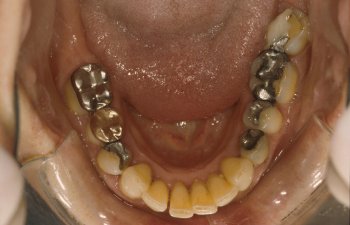
An often overlooked, but extremely important source of toxic material is the mercury from silver [mercury] amalgam fillings. Some people who are aware of the situation are confused by the mixture of information available. Unfortunately, statements from dental trade organizations and on a few poorly-researched news reports have muddled the situation.

Here are a few facts about mercury amalgam fillings:
Causes Damage to Brain in ChildrenIn February, 1998, a group of the world's top mercury researchers announced that mercury from amalgam fillings can permanently damage the brain, kidneys, and immune system of children.

Amalgam Fillings Linked to Neurological Problems, Gastrointestinal Problems
The first large-scale epidemiological study of mercury and adverse reactions was recently completed and showed that of the symptoms looked at, there was a link seen to gastrointestinal problems, sleep disturbances, concentration problems, memory disturbances, lack of initiative, restlessness, bleeding gums and other mouth disorders.

Mercury / Alzheimer's Disease Connection Found
A study related to mercury and Alzheimer's Disease was recently completed by a team of scientists led by well-respected researcher Dr. Boyd Haley. They exposed rats to levels of mercury vapor diluted to account for size differences between humans and rats. The rats developed tissue damage "indistinguishable" from that of Alzheimer's Disease.

Repeating the experiment showed the same results. Dr. Haley is quoted as saying "I'm getting the rest of my fillings taken out right now, and I've asked my wife to have hers replaced too." Also see: http://www.holistic-dentistry.com/artalzeimer.asp

Amalgam Fillings Since 1970s Unstable
The type of mercury fillings that began to be used during the last couple of decades, non-gamma-2 (high copper), releases many times more mercury than the older style of amalgam fillings. Also, please see the article on the instability of dental amalgam fillings by Ulf Bengsston.

Amalgam Fillings Release Highly Toxic Elemental MercuryMercury is one of the most toxic substances known. The mercury release from fillings is absorbed primarily as highly toxic elemental mercury vapor.

Amalgam Fillings Largest Source of Mercury
By FarBased on a number of studies in Sweden, the World Health Organization review of inorganic mercury in 1991 determined that mercury absorption is estimated to be approximately four times higher from amalgam fillings than from fish consumption. Recent studies have confirmed this estimate. The amount absorbed can vary considerably from person to person.

Gold Crowns, Gum, Bruxism, Computer Monitors Increase Release of Mercury Significantly
Gum chewing, grinding of teeth/bruxism, computer terminal exposure, presence of gold fillings or gold crowns (even if covering mercury fillings), teeth brushing, braces, and chewing cause the release of significantly increased amounts of mercury from the fillings.

Also, please see the following short review related to increases in mercury exposure from dissimilar metals in the mouth, exposure to magnetic fields, chewing, etc.

Cumulative Poison and Builds Up in OrgansMercury released from fillings builds up in the brain, pituitary, adrenals, and other parts of the body.

Mercury Amalgam Fillings Effect Porphyrins
Preliminary results from the first detailed biochemical analysis (scroll half-way down) of patients who removed mercury amalgam fillings showed a significant drop in the excretion of porphyrins (important to heme synthesis -- heme carries oxygen to red blood cells), as well as a number of other key biochemical changes. Also, see the Video of the preliminary results from the study.

Potential Contribuatory Factor in Other Diseases
Mercury from amalgam fillings has been implicated as a possible contribuatory factor in some cases of multiple sclerosis, Parkinson's Disease, IBS, reproductive disorders, allergies, and a variety of other illnesses.

Mercury Build Up in Brain, Organs and Breast Milk of Fetuses of Mothers With Amalgam FillingsMercury from fillings in pregnant women has been shown to cause mercury accumulation in brain, kidneys and liver of human fetuses (all of the areas tested). Studies have shown that mercury can be passed to infants from breast milk.

Proper Removal of Fillings Produces Eventual Health Improvement
A recent study published in the Journal of Orthomolecular Medicine related to the proper removal of mercury amalgam fillings from 118 subjects showed an elimination or reduction or 80% of the classic mercury poisoning symptoms. In many cases, it took 6 to 12 months after mercury amalgam removal for the symptoms to disappear.

World-reknowned Experts Agree About Potential Danger
In contrast to statements from dental trade organizations, toxicologists and medical researchers are often quite concerned about the use of mercury. Lars Friberg, the lead toxicologist on the World Health Organization team looking at inorganic mercury and health effects recently stated that he believes that mercury is unsuitable for dental materials because of safety concerns.

Canadian Class Action LawsuitCanadians are in the process of beginning a major class action lawsuit based on the fact that the government knew of but did not warn the public about mercury dangers from amalgam fillings. Legal actions related to mercury exposure from mercury amalgam fillings and vaccines are beginning in the United States. For more information and a directory of Mercury-free dentists, please see the TalkInternation.com web site.

Obviously, not everyone experiences acute toxicity effects from the mercury in amalgam fillings. However, virtually everyone does have mercury build up in their bodies from implantation of such fillings. The large increase in mercury exposure from the newer non-gamma-2 mercury fillings means that only time will tell how much damage has been caused by daily exposure to mercury to such fillings.I do not recommend that people assume automatically that they will be healed by the removal of amalgam fillings.

Many people are helped tremendously and some are healed. The 80% figure for people showing improvement within a year likely refers to people who had good reason to suspect that they were being significantly effected by the fillings. The percentage of people in the general population who might experience health improvement within one year after removal is probably much lower than 80%.

I recommend going into the mercury amalgam removal procedure knowing that, at the very least, you will have removed yourself from a regular exposure to an extremely toxic material such that it will not build up in your organs and possibly cause significant health problems at a later date.Mercury amalgam fillings should be removed only by dentists with experience using the IOAMT mercury amalgam removal protocol (presented with the permission of the excellent Preventive Dental Association web page).

Such dentists are often experienced with proper evaluation and placement of composite fillings, both of which can be crucial for the success of the treatment. Biocompatability tests are often important in determining which composite materials can be safely used. I believe that composite (plastic) fillings are a better replacement than metal (e.g., gold) fillings even in chemically-sensitive individuals.

They are, however, not without safety questions, but are still likely to be much less toxic than mercury amalgam fillings. Proper placement of composites should be left to experienced amalgam removal dentists as the average well-meaning dentist may not be aware of the newer placement techniques.

Further scientific information can be found at Mercury Adverse Effects Web Page, 150 Year's of Russian Roulette Web Page, Alt Corp's Amalgam Page, and Bo Walhjalt's Mercury Articles Web Page. More information about removal, detoxification, and placement of composite fillings can be found at Bioprobe, Inc. and at the Preventive Dental Association. Information about finding a dentist practicing non-toxic dentistry can be found on the Resources For Related to Non-Toxic Dentistry web page.

Also, the AMALGAM mailing list can be a good source of accurate, up-to-date information.

Important Links

Saturday, December 23, 2006

MERCURY IS A DEADLY POISON!

It is surprising how the controversy regarding mercury still continues even though the research indicating that it is a lethal poison is accumulating rapidly.

This research has shown how detrimental mercury can be to the developing foetus, the newborn, the developing child and the adult.

My intention here is to present a few research studies that will show the various detrimental effects of mercury at all stages of life.


Is there a correlation between the number of amalgams and the amount of mercury excreted in the urine after provocation?

ABSTRACT:

There is a considerable controversy as to whether dental amalgams may cause systemic health effects in humans because they liberate elemental mercury. Most such amalgams contain as much as 50% metallic mercury.

To determine the influence of dental amalgams on the mercury body burden of humans, we have given volunteers, with and without amalgams in their mouth, the sodium salt of 2, 3-dimercaptopropane-1-sulfonic acid (DMPS), a chelating agent safely used in the Soviet Union and West Germany for a number of years. The diameters of dental amalgams of the subjects were determined to obtain the amalgam score.

Administration of 300 mg DMPS by mouth increased the mean urinary mercury excretion of the amalgam group from 0.70 to 17.2 ug and that of the non amalgam group from 0.27 to 5.1 ug over a 9 hour period.

Two-thirds of the mercury excreted in the urine of those with dental amalgams appears to be derived originally from the mercury vapor released from their amalgams.

Linear regression analysis indicated a highly significant positive correlation between the mercury excreted in the urine 2 hours after DMPS administration and the dental amalgam scores. DMPS can be used to increase the urinary excretion of mercury and thus increase the significance and reliability of this measure of mercury exposure or burden, especially in cases of micromercurialism.

Aposhian, H.V., D.C. Bruce, W. Alter, R.C. Dart, K.M. Hurlbut, M.M. Aposhian, "Urinary Mercury after Administration of 2, 3-dimercaptopropane-1-sulfonic acid: Correlation with Dental Amalgam Score" FASEB J. 6: 2472-2476; (1992).

Can dental mercury release from the mother be detected in the foetus?

ABSTRACT:
In humans, the continuous release of Hg vapour from dental amalgam tooth restorations is markedly increased for prolonged periods after chewing. The present study establishes a time-course distribution for amalgam, Hg in body tissues of adult and foetal sheep. Under general anaesthesia, five pregnant ewes had twelve occlusal amalgam fillings containing radioactive 203Hg placed in teeth at 112 days gestation.

Blood, amniotic fluid, faeces, and urine specimens were collected at 1- to 3-day intervals for 16 days. From days 16-140 after amalgam placement (16-41 days for foetal lambs), tissue specimens were analyzed for radioactivity, and total Hg concentrations were calculated. Results demonstrate that Hg from dental amalgam will appear in maternal and foetal blood and amniotic fluid within 2 days after placement of amalgam tooth restorations.

Excretion of some of this Hg will also commence within 2 days. All tissues examined displayed Hg accumulation. Highest concentrations of Hg from amalgam in the adult occurred in kidney and liver, whereas in the foetus the highest amalgam Hg concentrations appeared in the liver and pituitary glands. The placenta progressively concentrated Hg as gestation advanced to term, and milk concentration of amalgam Hg postpartum provides a potential source of Hg exposure to the newborn. It is concluded that accumulation of amalgam Hg progresses in maternal and foetal tissues to a steady state with advancing gestation and is maintained.Vimy, M.J., Y. Takahashi, and F.L. Lorscheider "Maternal-foetal distribution of mercury (203Hg) released from dental amalgam fillings." Am. J. Physiol. 258 (Regulatory Integrative Comp. Physiol. 27): R939-R945 (1990).

Can in utero exposure to mercury cause behavioural disturbances?

ABSTRACT:

Pregnant rats were either 1) administered methyl mercury (MeHg) by gavage, 2 mg/kg/day during days 6-9 of gestation, 2) exposed by inhalation to metallic mercury (Hg) vapour (1.8 mg/m3 air for 1.5 hours per day) during gestation days 14-19, 3) exposed to both MeHg by gavage and Hg vapour by inhalation (MeHg + Hg), or 4) were given combined vehicle administration for each of the two treatments (control).

The inhalation regimen corresponded to an approximate dose of 0.1 mg Hg/kg/day.Clinical observations and developmental markers up to weaning showed no differences between any of the groups. Testing of behavioural functions was performed between 4 and 5 months of age and included spontaneous motor activity, spatial learning in a circular path, and instrumental maze learning for food reward.

Offspring of dams exposed to Hg vapour showed hyperactivity in the motor activity test chambers over all three parameters: locomotion, rearing and total activity; this effect was potentiated in the animals of the MeHg + Hg group. In the swim maze test, the MeHg + Hg and Hg groups evidenced longer latencies to reach a submerged platform, which they had learned to mount the day before, compared to either the control or MeHg group.

In the modified, enclosed radial arm maze, both the MeHg + Hg and Hg groups showed more ambulations and rearings in the activity test prior to the learning test. During the learning trial, the same groups (i.e., MeHg + Hg and Hg) showed longer latencies and made more errors in acquiring all eight pellets.Fredriksson, A., Dencker, L., Archer, T., Danielsson, B.R. "Prenatal Coexposure to Metallic Mercury Vapor and Methyl Mercury Produce Interactive Behavioral Changes in Adult Rats." Neurotoxicol Teratol., 18(2): 129-34, (1996).

ABSTRACT: The total mercury concentrations in the liver (Hg-L), the kidney cortex (Hg-K) and the cerebral cortex (Hg-C) of 108 children aged 1 day- 5 years, and the Hg-K and Hg-L of 46 foetuses were determined. As far as possible, the mothers were interviewed and their dental status was recorded.

The results were compared to mercury concentrations in the tissues of adults for the same geographical area. The Hg-K (n=38) and Hg-L (n=40) of foetuses and Hg-K (n=35) and Hg-C (n=35) of older infants (11-50 weeks of life) correlated significantly with the number of dental amalgam fillings of the mother. The toxicological relevance of the unexpected high Hg-K of older infants from mother with higher numbers of dental amalgam fillings is discussed. Conclusion: Future discussion on the pros and cons of dental amalgam should not be limited to adults or children with their own amalgam fillings, but also include foetal exposure.

The unrestricted application of amalgam for dental restorations in women before and during the child-bearing age should be reconsidered. Abbreviations: Hg-C total mercury concentration in the cerebral cortex (ng/g wet weight). Hg-K total mercury concentration in the renal cortex (ng/g wet weight). Hg-L total mercury concentration in the liver (ng/g wet weight).Drasch et. al. "Mercury Burden of Human Fetal and Infant Tissues" European Journal of Pediatrics (August 1994).

Can mercury amalgam from lactating mothers affect the foetus in utero?

ABSTRACT: Neonatal uptake of Hg from milk was examined in a pregnant sheep model, where radioactive mercury (Hg203)/silver tooth fillings (amalgam) were newly placed. A crossover experimental design was used in which lactating ewes nursed foster lambs. In a parallel study, the relationship between dental history and breast milk concentration of Hg was also examined.Results from the animal studies showed that, during pregnancy, a primary fetal site of amalgam, Hg concentration is in the liver, and after delivery the neonatal lamb kidney receives additional amalgam Hg from mother's milk.

In lactating women with aged amalgam fillings, increased Hg excretion in breast milk and urine correlated with the number of fillings or Hg vapor concentration levels in mouth air.It was concluded that Hg originating from maternal amalgam tooth fillings transfers across the placenta to the fetus, across the mammary gland into milk ingested by the newborn and ultimately into neonatal body tissues.

Comparisons are made to the U.S. minimal risk level recently established for adult Hg exposure. These findings suggest the placement and removal of "silver" tooth filings in pregnant and lactating humans will subject the fetus and neonate to unnecessary risk of Hg exposure.Vimy, M.J., Hooper, D.E., King, W.W., Lorscheider, F.L., "Mercury from Maternal "Silver" Tooth Fillings in Sheep and Human Breast Milk: A Source of Neonatal Exposure" Biological Trace Element Research, 56:143-52, (1997).

Can heavy metals affect human fertility?

ABSTRACT: Heavy metals have been identified as factors affecting human fertility. This study was designed to investigate whether the urinary heavy metal excretion is associated with different factors of infertility.

The urinary heavy metal excretion was determined in 501 infertile women after oral administration of the chelating agent 2,3-dimercaptopropane-1-sulfonic acid (DMPS). Furthermore, the influence of trace element and vitamin administration on metal excretion was investigated. Significant correlations were found between different heavy metals and clinical parameters (age, body mass index, nationality) as well as gynaecological conditions (uterine fibroids, miscarriages, hormonal disorders).

Diagnosis and reduction of an increased heavy metal body load improved the spontaneous conception chances of infertile women. The DMPS test was a useful and complementary diagnostic method. Adequate treatment provides successful alternatives to conventional hormonal therapy.Gerhard, I., Monga, B., Waldbrenner, A., Runnebaum, B., "Heavy Metals and Fertility" Journal of Toxicology and Environmental Health, Part, A, 54:593-611, (1998).

Is mercury associated with cardiac dysfunction?

OBJECTIVES: We sought to investigate the possible pathogenetic role of myocardial trace elements (TE) in patients with various forms of cardiac failure.BACKGROUND: Both myocardial TE accumulation and deficiency have been associated with the development of heart failure indistinguishable from an idiopathic dilated cardiomyopathy.

METHODS: Myocardial and muscular content of 32 TE has been assessed in biopsy samples of 13 patients (pts) with clinical, hemodynamic and histologic diagnosis of idiopathic dilated cardiomyopathy (IDCM), all without past or current exposure to TE.

One muscular and one left ventricular (LV) endomyocardial specimen from each patient, drawn with metal contamination-free technique, were analyzed by neutron activation analysis and compared with

1) similar surgical samples from patients with valvular (12 pts)and ischemic (13 pts) heart disease comparable for age and degree of LV dysfunction;

2) papillary and skeletal muscle surgical biopsies from 10 pts with mitral stenosis and normal LV function, and

3) LV endomyocardial biopsies from four normal subjects.

RESULTS: A large increase (>10,000 times for mercury and antimony) of TE concentration has been observed in myocardial but not in muscular samples in all pts with IDCM.

Patients with secondary cardiac dysfunction had mild increase (< or = 5 times) of myocardial TE and normal muscular TE. In particular, in pts with IDCM mean mercury concentration was 22,000 times (178,400 ng/g vs. 8 ng/g), antimony 12,000 times (19,260 ng/g vs. 1.5 ng/g), gold 11 times (26 ng/g vs. 2.3 ng/g), chromium 13 times (2,300 ng/g vs. 177 ng/g) and cobalt 4 times (86,5 ng/g vs. 20 ng/g) higher than in control subjects.

CONCLUSIONS: A large, significant increase of myocardial TE is present in IDCM but not in secondary cardiac dysfunction. The increased concentration of TE in pts with IDCM may adversely affect mitochondrial activity and myocardial metabolism and worsen cellular function.Frustaci A, Magnavita N, Chimenti C, Caldarulo M, Sabbioni E, Pietra R, Cellini C, Possati GF, Maseri A. Department of Cardiology, Catholic University, Rome, Italy. "Marked elevation of myocardial trace elements in idiopathic dilated cardiomyopathy compared with secondary cardiac dysfunction." From: J Am Coll Cardiol 1999 May;33(6):1578-83

Can dental mercury provoke an increase in antibiotic-resistant bacteria in oral and intestinal flora?

ABSTRACT: In a survey of 640 human subjects, a subgroup of 356 persons without recent exposure to antibiotics demonstrated that those with a high prevalence of Hg resistance in their intestinal floras were significantly more likely to also have resistance to two or more antibiotics. This observation led us to consider the possibility that mercury released from amalgam ("silver") dental restorations might be a selective agent for both mercury- and antibiotic-resistant bacteria in the oral and intestinal floras of primates.

Resistances to mercury and the several antibiotics were examined in the oral and intestinal floras of six adult monkeys prior the installation of amalgam fillings, during the time they were in place, and after replacement of the amalgam fillings with glass ionomer fillings (in four of the monkeys). The monkeys were fed an antibiotic-free diet, and fecal mercury concentrations were monitored.

There was a statistically significant increase in the incidence of mercury-resistant bacteria during the 5 weeks following installation of the amalgam fillings and during the 5 weeks immediately following their replacement with glass ionomer fillings. These peaks in incidence of mercury-resistant bacteria correlated with peaks of Hg elimination (as high as 1mM in the faeces) immediately following amalgam placement and immediately after replacement of the amalgam fillings.

Representative mercury-resistant isolates of three selected bacterial families (oral streptococci, members of the family Enterobacteriaceae, and enterocaocci) were also resistant to one or more antibiotics, including ampicillin, tetracycline, streptomycin, kanamycin, and chloramphenicol. While such mercury- and antibiotic-resistant isolates among the staphylococci, the enterococci, and members of the family Enterobacteriaceae, have been described, this is the first report of mercury resistance in the oral streptococci.

Many of the enterobacterial strains were able to transfer mercury and antibiotic resistances together to laboratory bacterial recipients, suggesting that the loci for these resistances are genetically linked.

Our findings indicate that mercury released from amalgam fillings can cause an enrichment of mercury resistance plasmids in the normal bacterial floras of primates. Many of these plasmids also carry antibiotic resistance, implicating the exposure to mercury from dental amalgams in an increased incidence of multiple antibiotic resistance plasmids in the normal floras of nonmedicated subjects.

Summers, A.O., J.Wireman, M.J. Vimy, F.L. Lorscheider, B. Marshall, S.B. Levy, S. Bennett, and L. Billard, "Mercury Released form Dental "Silver" Fillings Provokes an Increase in Mercury- and Antibiotic-Resistant Bacteria in Oral and Intestinal Floras of Primates", Antimicrobial Agents and Chemotherapy, (April 1993), pages 825 - 834.

Are there increased blood mercury levels in patients with Alzheimer's Disease?

SUMMARY: Alzheimer's disease (AD) is a common neurodegenerative disorder that leads to dementia and death. In addition to several genetic parameters, various environmental factors may influence the risk of getting AD.

In order to test whether blood levels of the heavy metal mercury are increased in AD, we measured blood mercury concentrations in AD patients (n=33), and compared them to age-matched control patients with major depression (MD) (n=45), as well as to an additional control group of patients with various non psychiatric disorders (n=65). Blood mercury levels were more than two fold higher in AD patients as compared to both control groups (p=0.0005, and p=0.0000, respectively). In early onset AD patients (n=13), blood mercury levels were almost three fold higher as compared to controls (p=0.0002, and p=0.0000, respectively).

These increases were unrelated to the patients' dental status. Linear regression analysis of blood mercury concentrations and CSF levels of amyloid B-peptide (AB) revealed a significant correlation of these measures in AD patients (n=15, r=0.7440, p=0.0015, Pearson type of correlation).

These results demonstrate elevated blood levels of mercury in AD, and they suggest that this increase of mercury levels is associated with high CSF levels of AB, whereas tau levels were unrelated. Possible explanations of increased blood mercury levels in AD include yet unidentified environmental sources or release from brain tissue with the advance in neuronal death.C. Hock, G. Drasch, S. Golombowski, F. Muller-Spahn, B. Willershausen-Zonnchen, P. Schwarz, U. Hock, J.H. Growdon, R.M. Nitsch "Increased Blood Mercury Levels in Patients with Alzheimer's Disease" Journal of Neural Transmission, 105: (1998).

Chemical & Heavy Metal Cleanse Starter Kit

Chemical & Heavy Metal Cleanse Starter Kit

$149.85
[ learn more ]

Add to Cart

The Chemical & Heavy Metal Starter Kit was designed by Dr. Group for individuals that are new to the cleansing process, or are simply looking for an easy-to-perform, cost effective cleanse program. The Heavy Metal Starter Kit is comprised of LIFE Detox Foot Patches™, NDF Plus™, and Quantum Zeolite™.

Toxicity of amalgam

It is surprising how the controversy regarding mercury still continues, even though the research indicating that it is a lethal poison, is accumulating rapidly. This research has shown how detrimental mercury can be to the developing foetus, the newborn, the developing child and the adult. My intention here is to present a few research studies that will show the various detrimental effects of mercury at all stages of life.

The major topics covered will include:

  • Is there a correlation between the number of amalgams and the amount of mercury excreted in the urine after provocation?
  • Can dental mercury released from the mother be detected in the foetus?
  • Can in utero exposure to mercury cause behavioural disturbances?
  • Can mercury amalgam from lactating mothers affect the foetus in utero?
  • Can heavy metals affect human fertility?
  • Is mercury associated with cardiac dysfunction?
  • Can dental mercury provoke an increase in antibiotic-resistant bacteria in oral and intestinal flora?
  • Are there increased blood mercury levels in patients with Alzheimer's disease?

    more information at:
  • detoxmetals
  • chelation.htm

more discussion: Forum
· Addiction Forum · Ask the Doctors Forum · Ayurveda Forum · Ayurvedic & Thai Herbs Forum · Colon Cleansing Forum · Dental Forum · Diabetes Forum · Diet Forum · General Cleansing Forum · Hepatitis A, B. C Forum · Integrated Medicine Forum · Live Blood Analysis Forum · Ozone-Oxygen-Forum · pH - Alkaline - Acidity Forum · Weight Loss Forum

Tuesday, November 14, 2006

What is EDTA chelation therapy and what is it used for?

What is EDTA chelation therapy and what is it used for?

Chelation (pronounced key-lay-shun) is the process by which a metal or mineral (such as lead, mercury, iron, arsenic, aluminum, etc.) is bonded to another substance-in this case an amino acid called EDTA, Ethylene-Diamine-Tetra-Acetic acid.

It is a natural process, basic to life itself. During EDTA chelation therapy, the EDTA infusion bonds with unwanted metals in the body and quickly carries them away in the urine.

Chelation therapy is a safe, effective alternative to drugs and surgeries and is used to treat many illnesses now known to be linked to the presence of toxic heavy metals.

Illnesses such as heart disease, strokes, diabetes, circulatory disorders, neuropathies, Alzheimer's disease, atherosclerosis, and adverse reactions to many environmental pollutants.

Traditional chelation therapy uses an intravenous drip, and is administered in the outpatient setting. The number of treatments vary based on each person's individual condition and/or goals of treatment. The average therapy is given one to three times a week for twenty to thirty treatments.

How long has EDTA chelation therapy been in use? Why don't more people use it?

EDTA chelation therapy for the detoxification for heavy metals has been in continuous use since the 1940s when it was introduced specifically for the treatment of lead poisoning.

It was very quickly observed that as the metals were eliminated, not only did the signs and symptoms of lead poisoning abate, but problems related to the circulatory system like heart attacks, angina, strokes, and peripheral vascular disease also improved.

For the past 50 years, well over one million people have received the intravenous form of EDTA chelation. As beneficial and life saving as this therapy has become, it is very expensive and very time-consuming, making it out of reach for most people.

Why is Ca-EDTA, Calcium Disodium EDTA, so much better than other types of chelation therapy?According to Dr. Bruce Halstead, "The chemistry of all chelators is such that a change of pH can dramatically effect the process of chemical binding needed to chelate a mineral or metal. When you use a less effective chelator, such as Magnesium EDTA, you lose all chelating ability of the two most essential heavy metals: lead and mercury.

Magnesium di-Potassium EDTA has a dramatically lower chelating effectiveness than Calcium EDTA because both magnesium and potassium dramatically decrease the pH in the blood environment to which it is introduced. Any factor decreasing pH renders EDTA less effective. Once the pH is lowered more than 7.38, it's no longer chemically conducive to any bonding or chelating." (Dr. Halstead is well known as the 'Father of Chelation Therapy'.)

Dr. Morton Walker Speaks on Detoxamin - Toxic Metals Induce Degenerative Diseases; Rectal Chelation Therapy Overcomes Them.

Environmentalists warn us repeatedly that we live on a poisoned planet. Toxins from mercury, lead, aluminum, cadmium, iron, nickel, and about 20 more metallic minerals permeate the Earth's milieu. Heavy and light metals poison us by combining to create deleterious signs and symptoms often referred to collectively as Toxic Metal Syndrome.

This syndrome, an indicator of serious systemic pathology, results in degenerative diseases which affect no less than 92% of the populations of Western industrialized nations, in particular, those people living in apartment high-rises and other polluted city dwellings.

What happens to them? These poisoned people eventually come down with manifestations of degenerative illnesses such as heart and/or blood vessel deteriorations; pancreatitis; gout, rheumatoid arthritis or osteoarthritis; the syndromes of yeast, chronic fatigue, and/or irritable bowel; Alzheimer's disease, multiple sclerosis, parkinsonism, and many more which may be deadly-cancer for instance.

Although a poisoned person's bones remain toxic for life, excellent self-treatment exists to reduce or reverse most symptoms of illness in other body parts.

First, get tested for the extent of toxicity, then neutralize metallic poisoning with a chelating agent such as Detoxamin. By applying the highly efficacious Detoxamin suppository containing EDTA, you remove toxic metal from cells all over the body.

The self-administration is performed rectally before retiring so that as you sleep you are taking chelation therapy with EDTA. There's no need for intravenous infusions or quantities of nutritional supplements.

Rectal chelation therapy does the job of detoxifying in a low-cost, convenient manner; it's an effective way to effuse EDTA through the bowel's walls and into your blood stream to clean toxic metals from all body cells.

Do I need Ca-EDTA chelation therapy?

We find ourselves existing in a far more toxic and hostile environment than our bodies were designed to handle.

Experts have shown that almost every health problem-from learning disorders to cancer and heart disease-is aggravated by the approximate 1,000% increase in lead levels in our bones. In 1999, it was reliably reported that hearts with some form of disease have 20,000 times more toxic heavy metals than healthy hearts.

"Human exposure to heavy metals has risen dramatically in the last 50 years as a result of an exponential increase in the use of heavy metals in industrial processes and products." says Maile Pouls, Ph.D (Townsend Letter for Doctors and Patients, July 1999).

A recently concluded "Body Burden" study by New York's Mt. Sinai Hospital and the Environmental Working Group was reviewed by University of Oregon Professor Joseph Thornton: "It shows the universality of chemical contamination of people's bodies," Thornton said.

All the studies "confirm the general message that everybody in our society has these chemicals building up. Some people have it worse than others, but everyone has it. No one is clean anymore." (From Being Careful Can't Keep Chemicals Out of Your Body, Miami Herald, February 1, 2003.)

Today we know that about one out of every 2.5 Americans will get cancer. Ninety eight percent of cancer is caused by toxic chemicals.

When 50% of all men and 33% of all women living now will die of cancer, something is terribly wrong. (Mortality from cancer was reduced by 90% during an 18-year study of 59 patients treated with Calcium-EDTA. This and over 40 other studies prove the efficacy of Ca-EDTA, Calcium Disodium EDTA chelation therapy and Detoxamin.

We will all function better and live longer if we lower the overall burden of toxic metals within ourselves. If you eat or breathe, you will probably benefit greatly from chelation therapy.

Is Detoxamin safe for children?

Yes. In fact, Detoxamin case studies were conducted on lead poisoning in children. The study showed no significant increase in BUN or creatinine levels even in very young children.

Due to our lower dosage and time release formulation, no renal toxicity was encountered.
BEHAVIORAL, STRUCTURAL, FUNCTIONAL ABNORMALITIES ASSOCIATED WITH VARIOUS HEAVY METAL TOXINS.

Reference: Published in the August issue of Alternative & Complimentary Therapies (a magazine for doctors) and Published in Townsend Letter for Doctor's and Patients
Psychiatric Disturbances:

Social Deficits, Social withdrawal

Mercury
Repetitive, perseverative, stereotyped behaviors; OCD-typical behaviors

Mercury
Depression, mood swings, flat affect; impaired facial recognition

Arsenic, Copper, Lead, Mercury
Schizoid tendencies; hallucinations; delirium

Mercury
Irritability, aggressive behaviors, temper tantrums

Lead, Mercury
Suicidal Behaviors

Copper, Mercury
Sleep difficulties / disturbances

Lead, Mercury, Thallium
Chronic fatigue (CFS); weakness, malaise

Aluminum, Arsenic, Cadmium, Copper, Lead, Mercury, Thallium
Anorexia; symptoms reflecting eating disorders, loss of appetite/weight

Arsenic, Lead, Mercury
Anxiety; nervous tendencies

Thallium
Attentional problems (ADHD), lacks eye contact, impaired visual fixation

Lead, Mercury
Speech and Language Deficits:
Speech disorders

Aluminum, Mercury
Loss of speech, developmental problems with language

Mercury
Speech comprehension deficits

Mercury
Dysarthria; articulation problems; slurred speech, unintelligible speech

Mercury
Cognitive Impairments:
Mental retardation, borderline intelligence

Arsenic, Lead, Mercury
Uneven performance on IQ scores, low IQ scores

Copper, Lead
Poor concentration, attention deficits (ADHD, response inhibition

Aluminum, Lead
Poor memory (short term, verbal, and auditory)

Aluminum, Lead
Difficulties understanding abstract ideas; difficulty carrying out complex commands

X metals
Dementia; pre-senile and senile dementia

Aluminum
Stupor

Aluminum, Arsenic
Impaired reaction time; lower performance on timed tests

Lead
Sensory Abnormalities:
Abnormal Sensations in the mouth and extremities

Arsenic
Hearing loss, difficulty hearingArsenic, Lead, Mercury
Abnormal touch sensations; diminished touch sensations, aversion to touch

Arsenic
Blurred vision; sensitivity to light

Arsenic, Mercury
Motor Disorders:
Choreiform movements, myoclonal jerks, unusualpostures

Copper, Mercury
Difficulty walking, swallowing, talking

Copper, Mercury
Flapping, circling, rocking, toe walkingMercury
Problems with intentional movements or imitation

Mercury
Abnormal, gait/posture; incoordination, loss of balance; problems sitting, lying, crawling and walking

Mercury
Decreased locomotor activity

Aluminum, Arsenic
Convulsion; seizure

Aluminum, Arsenic, Copper, Lead, Mercury, Thallium
Physiological Impairment, Brain and Central Nervous System:
Neurofibrillary tangles

Aluminum
Neuritis, retrobulbar neuritis; neuropathy

Aluminum, Arsenic, Lead, Thallium
Encephalopathy

Aluminum, Arsenic, Lead, Thallium
Cerebrovascular disease

X metals
Alterations in nerve conduction velocityLead
Alterations in the spinal cord

Thallium
Accumulates in CNS structures

Aluminum, Mercury
Abnormal EEGs

Arsenic, Lead
Autonomic disturbances

Copper, Lead, Mercury, Thallium
Peripheral Nervous System:
Peripheral neuropathy

Arsenic, Mercury
Alterations in peripheral nerves

Arsenic
Loss of feeling/ numbness in the extremities; paresthesia

Arsenic, Mercury, Thallium
Gastrointestinal Tract:
Nausea, vomiting, diarrhea; loss of appetite

Arsenic, Mercury
Abdominal pain, stomach cramps; burning of the throat of the mouth

Arsenic, Copper, Lead, Mercury, Thallium
Esophagitis; gastroenteritis; colitis

Arsenic, Mercury, Thallium Cancers (colon, pancreatic, stomach, or rectal) Arsenic
Renal and Hepatic Impairment:
Hepatotoxicity; Liver dysfunction, damage

Arsenic, Copper, Thallium
Cirrhosis of the liver; hepatitis

Copper
Kidney disease; kidney failure

Arsenic, Lead, Mercury
Renal toxicity; tubular proteinosis

Arsenic, Copper, Lead
Kidney Damage, histological alterations

Arsenic, Lead
Cardiovascular System:
Blood vessel damage

Arsenic
Anemia; decreased red blood cell count

Arsenic, Copper
Hypertension; increased heart rate (tachycardia)

Arsenic, Copper, Lead, Thallium
Electrocardiac disorders, Peripheral vascular disease; cardiovascular disease, vascular collapse

Arsenic, Lead
Respiratory System:
Pulmonary Fibrosis

Aluminum, Arsenic
Pulmonary edema

X metals
Pneumonia, laryngitis, pharyngitis, bronchitis

Aluminum, Arsenic, Mercury
Restrictive airway disorders, asthmatic conditions, pneumoconiosis

Arsenic, Aluminum
Nasal ulcers, perforation of the nasal septum
X metals
Immune System:
Increased incidences of Asthma, autoimmune-like symptoms, & allergies

X metals
Inhibition of lymphocytes, T-cells, monocytes X metals
Immunosuppression

Lead
Decreased white blood cell countArsenic, Thallium
Reproductive System:
Genital abnormalities

Aluminum, Thallium
Disturbances in menstrual cycle; menstrual pains

Copper, Mercury
Birth defects; premature births; Spontaneous abortion

Arsenic, Lead, Mercury
Reproductive dysfunction

Arsenic, Aluminum
Other Physical Disturbances:
Hypotonia or hypertonia; decreased muscular strength

X metal
Rashes, contact dermatitis; eczema, itchy/irritating skin

Aluminum, Arsenic, Copper, Mercury
Muscle pain; headache; acrodynia; colic

Arsenic, Copper, Lead, Thallium
Alopecia (hair loss)

Thallium
Reference: Published in the August issue of Alternative & Complimentary Therapies (a magazine for doctors) and Published in Townsend Letter for Doctor's and Patients.

Detoxamin Usage Instructions: Detoxamin EDTA Suppositories are solid, bullet-shaped preparations designed for easy insertion into the anus (back passage).

Detoxamin is manufactured in a cocoa-butter base, a time-release agent (fatty acid base), and 750 mg of Calcium-Disodium EDTA. Detoxamin will dissolve at body temperature and will gradually spread over the lining of the lower bowel (rectum), where it is absorbed into the bloodstream. Detoxamin is designed to release 750 mg Calcium Disodium EDTA slowly, over an 80-minute period.

A. Detoxamin Protocol for More Severe Cases:
1. Take one suppository at night, prior to bedtime.
2. Take every night for up to 90 days. This will provide the medical equivalence of 30 IV Chelation treatments.
3. Take proper mineral/trace mineral/vitamin replacement every day.
4. Take all other suplementaion every day.

B. Detoxamin Protocol for Less Severe Cases/Anti-Aging/Prevention:
1. Take Detoxamin every OTHER night, prior to bedtime.
2. Take Detoxamin every other night for 180 days (90 suppositories). This will provide the medical equivalence of 30 IV chelation treatments.
3. Take proper mineral/trace mineral/vitamin replacement every day.
4. Take all other supplementation every day.

Detoxamin Protocol AFTER A or B is completed:
Your bones are toxic for life. Lead and other heavy metals are stored in the bones and get re-distributed into the bloodstream. Therefore, it is highly recommended to continue maintenance with Detoxamin, this provides the ultimate in Anti-aging benefits.

1. Take 5 Detoxamin suppositories over a 30-day period. This porvides medically equal to about 2 EDTA IV treatments.
2. Take porper mineral/trace mineral/vitamin replacement every day.
Note: Detoxamin is designed to be taken at night, however some patients and physicians prefer taking a suppository in the morning after evacuation. (Optional)

How to Use Detoxamin:
Insert Detoxamin suppositories at night, prior to bedtime.
Eat early in the evening, about 4 hours prior to bedtime. (Reduces any discomfort).
1. Go to the toilet and empty your bowels if necessary.
2. Wash your hands.
3. Remove the plastic wrapping from Detoxamin.
4. Either squat or lie on your side with one leg bent and the other staight.
5. Gently but firmly push the suppository into the rectum, FLAT end first until past the sphincter muscle. By inserting the flat end first opposed to the pointed end, the suppository will travel higher up in the rectum more easily. If necessarey moisten the suppository with a little water. Push it in far enough so it doesn't slip out.
6. Close your legs and sit or lay still for a few minutes.
7. Wash your hands again.
8. Try not to empty your bowels for at least 80 minutes.
9. It is optional to take Detoxamin in the morning, after evacuation.
STORAGE: Store Detoxamin in a cool dark place, but not in the fridge.

If Detoxamin suppository gets warm it may melt, put the fridge for a few minutes, this will return the suppository to its original state so it may be inserted.

Shelf Life: 2 Years

Chemical & Heavy Metal Cleanse Starter Kit

Chemical & Heavy Metal Cleanse Starter Kit

$149.85
[ learn more ]

Add to Cart

The Chemical & Heavy Metal Starter Kit was designed by Dr. Group for individuals that are new to the cleansing process, or are simply looking for an easy-to-perform, cost effective cleanse program. The Heavy Metal Starter Kit is comprised of LIFE Detox Foot Patches™, NDF Plus™, and Quantum Zeolite™.

Dr. Group's Secret to Health Kit

Dr. Group's Secret to Health Kit

$39.94
[ learn more ]

Add to Cart

Dr. Group's Secret to Health Kit offers simple at-home solutions for cleansing internally and externally thereby reducing toxins, restoring the body's natural healing process, and helping you achieve true health and happiness.

Advanced Body Cleansing Kit

Advanced Body Cleansing Kit

$147.75
[ learn more ]

Add to Cart

Advanced Body Cleansing Kit with Livatrex™, Oxy-Powder®, Latero-Flora™ and two bottles of ParaTrex®.