 It is surprising how the controversy regarding mercury still continues even though the research indicating that it is a lethal poison is accumulating rapidly.
This research has shown how detrimental mercury can be to the developing foetus, the newborn, the developing child and the adult.
My intention here is to present a few research studies that will show the various detrimental effects of mercury at all stages of life.
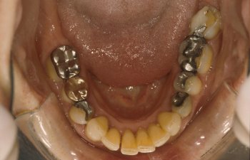
Is there a correlation between the number of amalgams and the amount of mercury excreted in the urine after provocation?
ABSTRACT:
There is a considerable controversy as to whether dental amalgams may cause systemic health effects in humans because they liberate elemental mercury. Most such amalgams contain as much as 50% metallic mercury.
To determine the influence of dental amalgams on the mercury body burden of humans, we have given volunteers, with and without amalgams in their mouth, the sodium salt of 2, 3-dimercaptopropane-1-sulfonic acid (DMPS), a chelating agent safely used in the Soviet Union and West Germany for a number of years. The diameters of dental amalgams of the subjects were determined to obtain the amalgam score.
Administration of 300 mg DMPS by mouth increased the mean urinary mercury excretion of the amalgam group from 0.70 to 17.2 ug and that of the non amalgam group from 0.27 to 5.1 ug over a 9 hour period.
Two-thirds of the mercury excreted in the urine of those with dental amalgams appears to be derived originally from the mercury vapor released from their amalgams.
Linear regression analysis indicated a highly significant positive correlation between the mercury excreted in the urine 2 hours after DMPS administration and the dental amalgam scores. DMPS can be used to increase the urinary excretion of mercury and thus increase the significance and reliability of this measure of mercury exposure or burden, especially in cases of micromercurialism.
Aposhian, H.V., D.C. Bruce, W. Alter, R.C. Dart, K.M. Hurlbut, M.M. Aposhian, "Urinary Mercury after Administration of 2, 3-dimercaptopropane-1-sulfonic acid: Correlation with Dental Amalgam Score" FASEB J. 6: 2472-2476; (1992).
Can dental mercury release from the mother be detected in the foetus?
ABSTRACT:
In humans, the continuous release of Hg vapour from dental amalgam tooth restorations is markedly increased for prolonged periods after chewing. The present study establishes a time-course distribution for amalgam, Hg in body tissues of adult and foetal sheep. Under general anaesthesia, five pregnant ewes had twelve occlusal amalgam fillings containing radioactive 203Hg placed in teeth at 112 days gestation.
Blood, amniotic fluid, faeces, and urine specimens were collected at 1- to 3-day intervals for 16 days. From days 16-140 after amalgam placement (16-41 days for foetal lambs), tissue specimens were analyzed for radioactivity, and total Hg concentrations were calculated. Results demonstrate that Hg from dental amalgam will appear in maternal and foetal blood and amniotic fluid within 2 days after placement of amalgam tooth restorations.
Excretion of some of this Hg will also commence within 2 days. All tissues examined displayed Hg accumulation. Highest concentrations of Hg from amalgam in the adult occurred in kidney and liver, whereas in the foetus the highest amalgam Hg concentrations appeared in the liver and pituitary glands. The placenta progressively concentrated Hg as gestation advanced to term, and milk concentration of amalgam Hg postpartum provides a potential source of Hg exposure to the newborn. It is concluded that accumulation of amalgam Hg progresses in maternal and foetal tissues to a steady state with advancing gestation and is maintained.Vimy, M.J., Y. Takahashi, and F.L. Lorscheider "Maternal-foetal distribution of mercury (203Hg) released from dental amalgam fillings." Am. J. Physiol. 258 (Regulatory Integrative Comp. Physiol. 27): R939-R945 (1990).
Can in utero exposure to mercury cause behavioural disturbances?
ABSTRACT:
Pregnant rats were either 1) administered methyl mercury (MeHg) by gavage, 2 mg/kg/day during days 6-9 of gestation, 2) exposed by inhalation to metallic mercury (Hg) vapour (1.8 mg/m3 air for 1.5 hours per day) during gestation days 14-19, 3) exposed to both MeHg by gavage and Hg vapour by inhalation (MeHg + Hg), or 4) were given combined vehicle administration for each of the two treatments (control).
The inhalation regimen corresponded to an approximate dose of 0.1 mg Hg/kg/day.Clinical observations and developmental markers up to weaning showed no differences between any of the groups. Testing of behavioural functions was performed between 4 and 5 months of age and included spontaneous motor activity, spatial learning in a circular path, and instrumental maze learning for food reward.
Offspring of dams exposed to Hg vapour showed hyperactivity in the motor activity test chambers over all three parameters: locomotion, rearing and total activity; this effect was potentiated in the animals of the MeHg + Hg group. In the swim maze test, the MeHg + Hg and Hg groups evidenced longer latencies to reach a submerged platform, which they had learned to mount the day before, compared to either the control or MeHg group.
In the modified, enclosed radial arm maze, both the MeHg + Hg and Hg groups showed more ambulations and rearings in the activity test prior to the learning test. During the learning trial, the same groups (i.e., MeHg + Hg and Hg) showed longer latencies and made more errors in acquiring all eight pellets.Fredriksson, A., Dencker, L., Archer, T., Danielsson, B.R. "Prenatal Coexposure to Metallic Mercury Vapor and Methyl Mercury Produce Interactive Behavioral Changes in Adult Rats." Neurotoxicol Teratol., 18(2): 129-34, (1996).
ABSTRACT: The total mercury concentrations in the liver (Hg-L), the kidney cortex (Hg-K) and the cerebral cortex (Hg-C) of 108 children aged 1 day- 5 years, and the Hg-K and Hg-L of 46 foetuses were determined. As far as possible, the mothers were interviewed and their dental status was recorded.
The results were compared to mercury concentrations in the tissues of adults for the same geographical area. The Hg-K (n=38) and Hg-L (n=40) of foetuses and Hg-K (n=35) and Hg-C (n=35) of older infants (11-50 weeks of life) correlated significantly with the number of dental amalgam fillings of the mother. The toxicological relevance of the unexpected high Hg-K of older infants from mother with higher numbers of dental amalgam fillings is discussed. Conclusion: Future discussion on the pros and cons of dental amalgam should not be limited to adults or children with their own amalgam fillings, but also include foetal exposure.
The unrestricted application of amalgam for dental restorations in women before and during the child-bearing age should be reconsidered. Abbreviations: Hg-C total mercury concentration in the cerebral cortex (ng/g wet weight). Hg-K total mercury concentration in the renal cortex (ng/g wet weight). Hg-L total mercury concentration in the liver (ng/g wet weight).Drasch et. al. "Mercury Burden of Human Fetal and Infant Tissues" European Journal of Pediatrics (August 1994).
Can mercury amalgam from lactating mothers affect the foetus in utero?
ABSTRACT: Neonatal uptake of Hg from milk was examined in a pregnant sheep model, where radioactive mercury (Hg203)/silver tooth fillings (amalgam) were newly placed. A crossover experimental design was used in which lactating ewes nursed foster lambs. In a parallel study, the relationship between dental history and breast milk concentration of Hg was also examined.Results from the animal studies showed that, during pregnancy, a primary fetal site of amalgam, Hg concentration is in the liver, and after delivery the neonatal lamb kidney receives additional amalgam Hg from mother's milk.
In lactating women with aged amalgam fillings, increased Hg excretion in breast milk and urine correlated with the number of fillings or Hg vapor concentration levels in mouth air.It was concluded that Hg originating from maternal amalgam tooth fillings transfers across the placenta to the fetus, across the mammary gland into milk ingested by the newborn and ultimately into neonatal body tissues.
Comparisons are made to the U.S. minimal risk level recently established for adult Hg exposure. These findings suggest the placement and removal of "silver" tooth filings in pregnant and lactating humans will subject the fetus and neonate to unnecessary risk of Hg exposure.Vimy, M.J., Hooper, D.E., King, W.W., Lorscheider, F.L., "Mercury from Maternal "Silver" Tooth Fillings in Sheep and Human Breast Milk: A Source of Neonatal Exposure" Biological Trace Element Research, 56:143-52, (1997).
Can heavy metals affect human fertility?
ABSTRACT: Heavy metals have been identified as factors affecting human fertility. This study was designed to investigate whether the urinary heavy metal excretion is associated with different factors of infertility.
The urinary heavy metal excretion was determined in 501 infertile women after oral administration of the chelating agent 2,3-dimercaptopropane-1-sulfonic acid (DMPS). Furthermore, the influence of trace element and vitamin administration on metal excretion was investigated. Significant correlations were found between different heavy metals and clinical parameters (age, body mass index, nationality) as well as gynaecological conditions (uterine fibroids, miscarriages, hormonal disorders).
Diagnosis and reduction of an increased heavy metal body load improved the spontaneous conception chances of infertile women. The DMPS test was a useful and complementary diagnostic method. Adequate treatment provides successful alternatives to conventional hormonal therapy.Gerhard, I., Monga, B., Waldbrenner, A., Runnebaum, B., "Heavy Metals and Fertility" Journal of Toxicology and Environmental Health, Part, A, 54:593-611, (1998).
Is mercury associated with cardiac dysfunction?
OBJECTIVES: We sought to investigate the possible pathogenetic role of myocardial trace elements (TE) in patients with various forms of cardiac failure.BACKGROUND: Both myocardial TE accumulation and deficiency have been associated with the development of heart failure indistinguishable from an idiopathic dilated cardiomyopathy.
METHODS: Myocardial and muscular content of 32 TE has been assessed in biopsy samples of 13 patients (pts) with clinical, hemodynamic and histologic diagnosis of idiopathic dilated cardiomyopathy (IDCM), all without past or current exposure to TE.
One muscular and one left ventricular (LV) endomyocardial specimen from each patient, drawn with metal contamination-free technique, were analyzed by neutron activation analysis and compared with
1) similar surgical samples from patients with valvular (12 pts)and ischemic (13 pts) heart disease comparable for age and degree of LV dysfunction;
2) papillary and skeletal muscle surgical biopsies from 10 pts with mitral stenosis and normal LV function, and
3) LV endomyocardial biopsies from four normal subjects.
RESULTS: A large increase (>10,000 times for mercury and antimony) of TE concentration has been observed in myocardial but not in muscular samples in all pts with IDCM.
Patients with secondary cardiac dysfunction had mild increase (< or = 5 times) of myocardial TE and normal muscular TE. In particular, in pts with IDCM mean mercury concentration was 22,000 times (178,400 ng/g vs. 8 ng/g), antimony 12,000 times (19,260 ng/g vs. 1.5 ng/g), gold 11 times (26 ng/g vs. 2.3 ng/g), chromium 13 times (2,300 ng/g vs. 177 ng/g) and cobalt 4 times (86,5 ng/g vs. 20 ng/g) higher than in control subjects.
CONCLUSIONS: A large, significant increase of myocardial TE is present in IDCM but not in secondary cardiac dysfunction. The increased concentration of TE in pts with IDCM may adversely affect mitochondrial activity and myocardial metabolism and worsen cellular function.Frustaci A, Magnavita N, Chimenti C, Caldarulo M, Sabbioni E, Pietra R, Cellini C, Possati GF, Maseri A. Department of Cardiology, Catholic University, Rome, Italy. "Marked elevation of myocardial trace elements in idiopathic dilated cardiomyopathy compared with secondary cardiac dysfunction." From: J Am Coll Cardiol 1999 May;33(6):1578-83
Can dental mercury provoke an increase in antibiotic-resistant bacteria in oral and intestinal flora?
ABSTRACT: In a survey of 640 human subjects, a subgroup of 356 persons without recent exposure to antibiotics demonstrated that those with a high prevalence of Hg resistance in their intestinal floras were significantly more likely to also have resistance to two or more antibiotics. This observation led us to consider the possibility that mercury released from amalgam ("silver") dental restorations might be a selective agent for both mercury- and antibiotic-resistant bacteria in the oral and intestinal floras of primates.
Resistances to mercury and the several antibiotics were examined in the oral and intestinal floras of six adult monkeys prior the installation of amalgam fillings, during the time they were in place, and after replacement of the amalgam fillings with glass ionomer fillings (in four of the monkeys). The monkeys were fed an antibiotic-free diet, and fecal mercury concentrations were monitored.
There was a statistically significant increase in the incidence of mercury-resistant bacteria during the 5 weeks following installation of the amalgam fillings and during the 5 weeks immediately following their replacement with glass ionomer fillings. These peaks in incidence of mercury-resistant bacteria correlated with peaks of Hg elimination (as high as 1mM in the faeces) immediately following amalgam placement and immediately after replacement of the amalgam fillings.
Representative mercury-resistant isolates of three selected bacterial families (oral streptococci, members of the family Enterobacteriaceae, and enterocaocci) were also resistant to one or more antibiotics, including ampicillin, tetracycline, streptomycin, kanamycin, and chloramphenicol. While such mercury- and antibiotic-resistant isolates among the staphylococci, the enterococci, and members of the family Enterobacteriaceae, have been described, this is the first report of mercury resistance in the oral streptococci.
Many of the enterobacterial strains were able to transfer mercury and antibiotic resistances together to laboratory bacterial recipients, suggesting that the loci for these resistances are genetically linked.
Our findings indicate that mercury released from amalgam fillings can cause an enrichment of mercury resistance plasmids in the normal bacterial floras of primates. Many of these plasmids also carry antibiotic resistance, implicating the exposure to mercury from dental amalgams in an increased incidence of multiple antibiotic resistance plasmids in the normal floras of nonmedicated subjects.
Summers, A.O., J.Wireman, M.J. Vimy, F.L. Lorscheider, B. Marshall, S.B. Levy, S. Bennett, and L. Billard, "Mercury Released form Dental "Silver" Fillings Provokes an Increase in Mercury- and Antibiotic-Resistant Bacteria in Oral and Intestinal Floras of Primates", Antimicrobial Agents and Chemotherapy, (April 1993), pages 825 - 834.
Are there increased blood mercury levels in patients with Alzheimer's Disease?
SUMMARY: Alzheimer's disease (AD) is a common neurodegenerative disorder that leads to dementia and death. In addition to several genetic parameters, various environmental factors may influence the risk of getting AD.
In order to test whether blood levels of the heavy metal mercury are increased in AD, we measured blood mercury concentrations in AD patients (n=33), and compared them to age-matched control patients with major depression (MD) (n=45), as well as to an additional control group of patients with various non psychiatric disorders (n=65). Blood mercury levels were more than two fold higher in AD patients as compared to both control groups (p=0.0005, and p=0.0000, respectively). In early onset AD patients (n=13), blood mercury levels were almost three fold higher as compared to controls (p=0.0002, and p=0.0000, respectively).
These increases were unrelated to the patients' dental status. Linear regression analysis of blood mercury concentrations and CSF levels of amyloid B-peptide (AB) revealed a significant correlation of these measures in AD patients (n=15, r=0.7440, p=0.0015, Pearson type of correlation).
These results demonstrate elevated blood levels of mercury in AD, and they suggest that this increase of mercury levels is associated with high CSF levels of AB, whereas tau levels were unrelated. Possible explanations of increased blood mercury levels in AD include yet unidentified environmental sources or release from brain tissue with the advance in neuronal death.C. Hock, G. Drasch, S. Golombowski, F. Muller-Spahn, B. Willershausen-Zonnchen, P. Schwarz, U. Hock, J.H. Growdon, R.M. Nitsch "Increased Blood Mercury Levels in Patients with Alzheimer's Disease" Journal of Neural Transmission, 105: (1998).
Chemical & Heavy Metal Cleanse Starter Kit$149.85  The Chemical & Heavy Metal Starter Kit was designed by Dr. Group for individuals that are new to the cleansing process, or are simply looking for an easy-to-perform, cost effective cleanse program. The Heavy Metal Starter Kit is comprised of LIFE Detox Foot Patches™, NDF Plus™, and Quantum Zeolite™. |
No comments:
Post a Comment